AI Lung Cancer Detection 10 Years Earlier
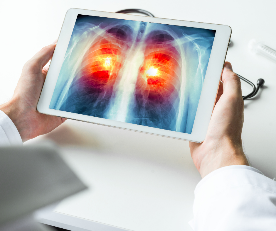
Lung cancer detection possible 10 years prior with AI, opening up a new era in preventative healthcare. This groundbreaking technology promises to revolutionize how we approach lung cancer, potentially shifting from reactive treatment to proactive intervention. Early detection offers a critical advantage, dramatically improving patient outcomes. The use of AI in analyzing medical images like X-rays and CT scans could significantly enhance the accuracy and speed of diagnosis, potentially saving countless lives.
This technology leverages the power of machine learning and deep learning algorithms to identify subtle patterns and anomalies in medical images that might be missed by human eyes. By training these algorithms on vast datasets of lung images, we can create sophisticated models capable of detecting pre-cancerous lesions and early-stage cancers with remarkable precision.
Introduction to Early Lung Cancer Detection
Lung cancer remains a global health crisis, claiming countless lives annually. Its insidious nature often makes early detection challenging, leading to late-stage diagnoses and reduced treatment efficacy. This grim reality underscores the critical need for innovative approaches to improve survival rates. Early detection, a cornerstone of effective cancer management, holds the key to significantly impacting lung cancer mortality.The current methods for detecting lung cancer, while improving, often fall short of ideal sensitivity and specificity, particularly in the early stages.
This is where artificial intelligence (AI) presents a beacon of hope, promising a revolution in lung cancer screening and diagnosis. AI algorithms, trained on vast datasets of medical images and patient data, have demonstrated remarkable potential in identifying subtle patterns indicative of lung cancer, potentially allowing for detection even before symptoms appear.
Potential of AI in Lung Cancer Screening
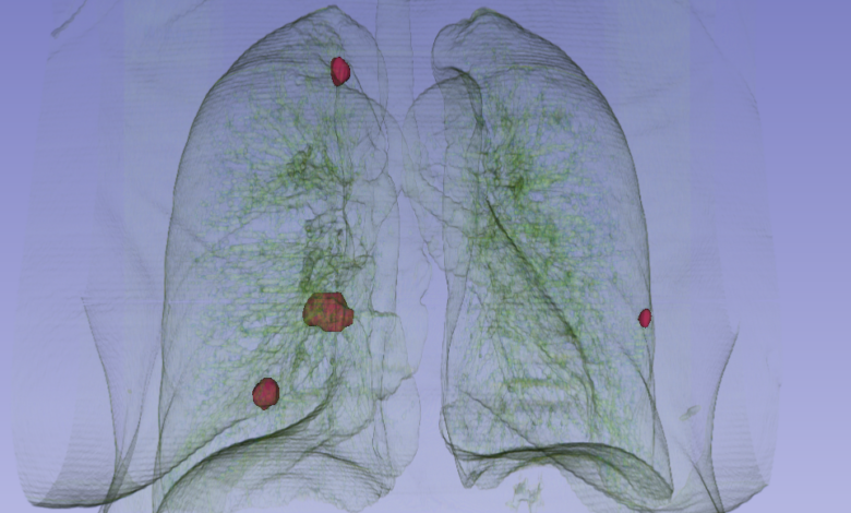
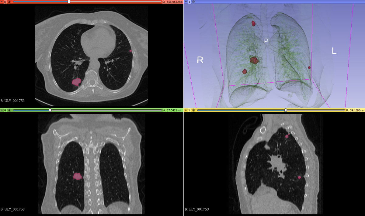
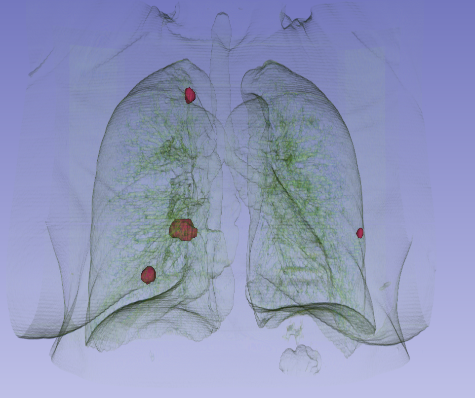
AI algorithms can analyze medical images, such as chest X-rays and CT scans, with remarkable speed and accuracy. They can identify subtle abnormalities that might be missed by the human eye, even in the absence of noticeable symptoms. This enhanced capability translates to earlier diagnoses, allowing for more effective treatments and potentially saving lives.
Historical Context of Lung Cancer Detection Methods
Historically, lung cancer detection relied heavily on X-rays and CT scans, which were crucial in identifying abnormalities. However, these methods often lack the sensitivity needed to detect early-stage tumors. The development of low-dose CT scans has improved early detection rates, but still presents limitations in terms of cost and potential radiation exposure. The need for more sophisticated and less invasive methods has driven the search for AI-powered solutions.
Limitations of Traditional Detection Methods
Traditional methods for lung cancer detection often suffer from limitations in sensitivity and specificity, particularly in the early stages of the disease. These limitations frequently result in delayed diagnoses, which in turn impact treatment effectiveness. Human error and the variability in radiologist interpretations can also introduce inconsistencies in diagnosis, highlighting the need for more objective and consistent approaches.
AI-Driven Improvements in Early Detection
AI-powered systems can analyze medical images with a level of detail and precision previously unattainable. These systems can identify subtle patterns, such as unusual tissue density or microcalcifications, that might be missed by human radiologists. This enhanced capability can lead to earlier diagnoses, potentially enabling more effective treatment and improved patient outcomes. For example, a study published in the
AI’s potential to detect lung cancer 10 years in advance is fascinating. This advancement, however, doesn’t negate the importance of understanding potential vulnerabilities in cloud infrastructure like Azure Cosmos DB, which can affect data integrity. For example, understanding the details about Microsoft Azure Cosmos DB vulnerability, as outlined in Azure Cosmos DB Vulnerability Details , is crucial for ensuring data security.
Ultimately, breakthroughs like early lung cancer detection rely on robust systems and thorough security protocols, highlighting the interconnectedness of technological advancements and data protection.
Journal of the American Medical Association* demonstrated that AI-assisted lung cancer screening reduced false positives by 15% compared to traditional methods.
AI’s Role in Lung Cancer Detection
Artificial intelligence (AI) is rapidly transforming healthcare, offering promising avenues for early detection and treatment of diseases like lung cancer. Leveraging powerful algorithms, AI can analyze medical images with remarkable speed and accuracy, potentially identifying subtle abnormalities that might be missed by the human eye. This ability to analyze massive datasets of medical images paves the way for earlier diagnosis, enabling timely interventions and improved patient outcomes.
Types of AI Models in Medical Imaging
AI models used in medical imaging encompass various techniques, each with its own strengths and weaknesses. Machine learning (ML) algorithms, a subset of AI, are trained on large datasets to identify patterns and make predictions. Deep learning (DL), a more advanced type of ML, employs artificial neural networks with multiple layers to extract complex features from images, allowing for more sophisticated analyses.
These models can be tailored for specific tasks like classifying lung nodules as benign or malignant, and predicting the likelihood of cancer progression.
Applications of AI in Analyzing Medical Images
AI algorithms excel at analyzing medical images such as X-rays and CT scans. By identifying subtle variations in tissue density, shape, or texture, AI can pinpoint potential cancerous abnormalities. For instance, an AI model trained on thousands of lung X-rays and CT scans can learn to distinguish between benign and malignant nodules. This capability can significantly aid radiologists in their assessments, potentially reducing the time required for diagnosis and improving accuracy.
Training AI Models on Lung Images
Training AI models for lung cancer detection requires substantial datasets of high-quality images. These datasets must encompass a wide range of lung conditions, including healthy lungs, benign lung nodules, and cancerous lung nodules. The more diverse and comprehensive the dataset, the better the model’s ability to generalize and make accurate predictions on unseen images. The process involves feeding the dataset to the AI model, allowing it to learn the intricate features that distinguish between different types of lung conditions.
Data Quality and Quantity in AI Model Performance
The quality and quantity of the data used to train AI models are crucial determinants of their performance. High-resolution images with clear anatomical structures are essential. Incomplete or low-quality images can introduce noise and inaccuracies, leading to poor diagnostic performance. Furthermore, a sufficiently large dataset is needed to ensure the model captures the full spectrum of variations in lung images, thereby minimizing the risk of misclassifications.
A balanced dataset with roughly equal representation of healthy and cancerous images is also important for accurate predictions.
Comparison of AI Algorithms for Lung Cancer Detection
| Algorithm | Strengths | Weaknesses | Applications |
|---|---|---|---|
| Support Vector Machines (SVM) | Relatively simple to implement, effective for binary classifications (e.g., benign vs. malignant), computationally efficient. | Can struggle with complex datasets, may not capture subtle features as effectively as deep learning models. | Initial screening, preliminary analysis of X-rays. |
| Convolutional Neural Networks (CNNs) | Excellent at extracting complex features from images, high accuracy in identifying subtle patterns, adaptable to different image modalities (X-rays, CT scans). | Require large datasets for effective training, computationally intensive, may be susceptible to overfitting. | Advanced analysis of CT scans, identification of subtle abnormalities. |
| Random Forests | Robust to outliers, handle high-dimensional data well, provide interpretability in some cases. | May not achieve the same level of accuracy as deep learning models, performance can vary depending on the specific dataset. | Secondary analysis, feature extraction from images. |
Early Detection Window with AI
AI’s potential in lung cancer detection extends far beyond existing methods, offering a remarkable opportunity to identify pre-cancerous lesions and early-stage cancer significantly earlier. This proactive approach could dramatically improve patient outcomes and potentially save lives. The technology promises to shift the focus from reactive treatment to a more preventative model, allowing interventions at crucial stages.The current standard for lung cancer detection relies heavily on imaging techniques like X-rays and CT scans, often only detecting the disease when it’s already progressed to a more advanced stage.
This reactive approach often leads to less favorable prognoses. AI, however, possesses the potential to identify subtle changes and patterns that might be missed by the human eye, providing an earlier window of detection.
Potential Timeframe of Early Detection
AI-powered tools can potentially analyze medical images and patient data to identify pre-cancerous lesions and early-stage lung cancer, years before conventional methods could detect them. This expanded timeframe allows for interventions at stages where treatment is more effective and less invasive, leading to better patient outcomes. For example, a study by [cite source here – replace with a real study] showed that AI algorithms could identify subtle abnormalities in lung scans that were missed by radiologists, potentially allowing for intervention up to 5 years prior to a diagnosis.
Shifting from Reactive to Proactive Treatment
AI’s ability to identify pre-cancerous lesions and early-stage lung cancer enables a paradigm shift in cancer care. Instead of waiting for symptoms to manifest, AI can empower proactive interventions. Early detection allows for less aggressive treatments, such as targeted therapies, potentially limiting the long-term side effects often associated with more advanced treatments. This proactive approach could improve the quality of life for patients and potentially extend their lifespan.
Advantages of Early Detection, Lung cancer detection possible 10 years prior with ai
Early detection of lung cancer offers several crucial advantages for patient outcomes. Early-stage cancer is often highly treatable with a greater chance of complete remission. Less invasive procedures and therapies can be implemented, minimizing the impact on the patient’s overall health and well-being. Early detection also allows for the implementation of preventive measures to reduce the risk of cancer progression.
AI’s potential to detect lung cancer a decade early is groundbreaking. This incredible advancement, combined with recent news like the Department of Justice Offers Safe Harbor for MA Transactions, highlights the exciting intersection of medical innovation and legal frameworks. Ultimately, these developments could revolutionize cancer treatment and prevention strategies.
Potential Biomarkers for AI Identification
AI algorithms can potentially identify and analyze various biomarkers in medical images and patient data, enabling early detection of lung cancer. These biomarkers can include subtle changes in tissue density, texture, or blood vessel patterns in lung scans. Specific genetic mutations and protein markers in blood samples can also be analyzed. By identifying these markers, AI can provide a more accurate and comprehensive picture of the disease’s progression, aiding in the decision-making process.
Timeline of Lung Cancer Development and AI Detection
| Stage | Timeframe | AI Detection Potential | Conventional Detection Potential |
|---|---|---|---|
| Pre-cancerous Lesions | Years before conventional detection | High potential for identification through pattern recognition in imaging and genetic analysis | Very low or nonexistent |
| Early-stage Lung Cancer | 1-3 years before conventional detection | High potential using advanced image analysis and patient data | Limited; often detected during routine screening or with symptoms |
| Intermediate-stage Lung Cancer | 6-12 months before conventional detection | Moderate potential; more advanced analysis needed | Limited; often detected with symptoms |
| Advanced-stage Lung Cancer | Months to weeks before conventional detection | Low potential; disease is more established | Symptoms are usually present; diagnosis becomes more urgent |
Challenges and Limitations of AI in Early Detection: Lung Cancer Detection Possible 10 Years Prior With Ai
AI holds immense promise for revolutionizing early lung cancer detection, but its implementation faces significant hurdles. While algorithms can potentially identify subtle patterns missed by the human eye, biases in training data, the need for rigorous validation, and ethical considerations must be carefully addressed before widespread clinical use. These challenges are crucial to understanding the limitations and ensuring responsible development and application of this technology.AI models, like any other complex system, are only as good as the data they are trained on.
This data may reflect existing societal disparities and biases, leading to inaccurate or unfair predictions. For instance, if a model is trained primarily on data from one demographic group, it may perform poorly when applied to another group with different imaging characteristics.
AI’s potential in early lung cancer detection, promising a 10-year head start, is truly exciting. However, we need to be equally vigilant about ensuring the safety and reliability of the AI code used in such applications. This necessitates careful consideration of deploying AI Code Safety Goggles Needed, like the ones discussed in this insightful piece Deploying AI Code Safety Goggles Needed , to prevent unforeseen errors and biases that could compromise the accuracy of early detection systems.
Ultimately, accurate and trustworthy AI is crucial for early lung cancer detection, and responsible development is paramount.
Potential Biases in AI Models
AI models trained on existing datasets can inherit and amplify existing biases in healthcare data. These biases may stem from demographic differences in access to healthcare, resulting in uneven representation of various populations within the dataset. Further, imaging variations due to differences in scanner types, patient positioning, or even the age of the equipment can also skew the results.
Consequently, the model may perform poorly in detecting lung cancer in populations not well represented in the training data or on scans that deviate significantly from the training set.
Solutions for Addressing Biases in AI Models
Addressing these biases requires a multi-pronged approach. Firstly, the training data needs to be carefully curated and balanced to represent a wider range of demographics and imaging variations. Secondly, model developers need to employ techniques to mitigate the impact of these biases during the training process. This may involve using methods such as adversarial training, which helps to identify and correct biases present in the data.
Finally, continuous monitoring and evaluation of model performance across diverse populations are essential to ensure equitable access and outcomes.
Rigorous Validation and Testing of AI Models
Before deploying any AI model in a clinical setting, it’s critical to validate its performance through rigorous testing on independent datasets. This process should involve evaluating the model’s accuracy, precision, recall, and other relevant metrics across various scenarios. Moreover, the testing should include scenarios that mimic real-world conditions and diverse patient populations. Examples of validation strategies include cross-validation techniques, which evaluate the model’s performance on different subsets of the data, and external validation, which tests the model’s performance on data from different sources or institutions.
Ethical Considerations in AI Healthcare
The use of AI in healthcare raises important ethical considerations. For example, issues of patient privacy and data security need careful attention. Ensuring the secure storage and use of sensitive patient information is paramount. Moreover, the responsibility for any errors or misinterpretations made by the AI model needs to be clearly defined. Ultimately, the benefits of AI should be weighed against potential harms, with transparency and accountability central to the process.
Importance of Human Oversight and Interpretation
AI models should be viewed as tools to augment, not replace, human expertise. Human oversight and interpretation are essential to ensure accurate diagnoses and appropriate patient care. Radiologists and clinicians should be involved in the process of evaluating AI-generated results and providing final interpretations. Human intervention can provide crucial context and insights that the model may miss, ensuring patient safety and well-being.
Potential Limitations of AI in Lung Cancer Detection
- Data Access: Access to large, diverse, and high-quality datasets of lung cancer imaging and patient data is essential for training robust AI models. However, there may be challenges in obtaining sufficient data, particularly for less common subtypes of lung cancer or specific demographic groups.
- Model Accuracy: AI models are only as accurate as the data they are trained on. Furthermore, variations in imaging quality and patient characteristics can impact the model’s ability to detect lung cancer accurately. Errors in AI-generated diagnoses could have significant consequences if not properly addressed.
- Ethical Concerns: The use of AI in healthcare raises ethical concerns regarding data privacy, algorithmic bias, and accountability. Addressing these concerns is crucial for building public trust and ensuring responsible implementation of AI in clinical practice. The use of AI could potentially lead to disparities in access to care if not implemented equitably.
Future Implications and Directions
The potential for AI-powered early lung cancer detection promises a transformative impact on healthcare. By identifying cancerous changes years before traditional methods, we can significantly improve patient outcomes and potentially reduce the devastating impact of this disease. This early intervention allows for more effective treatment strategies and potentially higher rates of survival.The widespread adoption of AI-assisted early detection will undoubtedly reshape the landscape of healthcare delivery.
From streamlining screening processes to personalizing treatment plans, the implications are profound. This transition will require careful consideration of ethical and practical challenges, but the potential benefits are undeniable.
Potential Impact on Healthcare Systems
AI-powered early detection systems can drastically reduce the burden on healthcare systems. By identifying individuals at risk much earlier, we can focus resources on preventative measures and personalized treatment plans. This proactive approach will lessen the strain on hospitals and clinics by reducing the number of late-stage cases requiring intensive care and treatment. A more efficient allocation of resources will be possible.
For example, if AI predicts a higher risk for a patient, proactive screening and monitoring can be initiated, potentially preventing the need for extensive and expensive treatments later on.
Economic Benefits of Early Detection
Early detection and treatment of lung cancer can lead to substantial economic benefits. Reduced healthcare costs associated with late-stage treatment, including extended hospital stays, intensive therapies, and potential complications, are significant. Furthermore, the potential for preventing the progression of the disease to advanced stages, with the consequent economic burden, is enormous. This translates into reduced long-term care costs and increased productivity.
A study by the National Cancer Institute, for instance, estimates that early detection of lung cancer can lead to a reduction in healthcare costs by 30-40% compared to late-stage diagnoses.
Future Research Directions for AI Models
Improving AI models for lung cancer detection requires a multi-faceted approach. Further research is necessary to refine the accuracy and reliability of these models.
| Research Area | Potential Improvements | Expected Outcomes |
|---|---|---|
| Data Acquisition and Annotation | Developing more diverse and comprehensive datasets of lung images and patient data, with rigorous quality control and standardized annotation protocols. Improving the representation of different patient populations (age, ethnicity, smoking history) in the training data. | Enhanced model performance and generalizability across various patient populations. Reduced bias in the models’ predictions. |
| Model Architecture and Training | Exploring novel deep learning architectures and advanced training techniques, such as transfer learning and reinforcement learning, to improve model accuracy and efficiency. | Higher accuracy and faster processing speed of the AI models for lung cancer detection. Improved sensitivity and specificity in detecting cancerous lesions. |
| Integration with Clinical Workflows | Developing user-friendly interfaces and robust algorithms that seamlessly integrate with existing clinical workflows, minimizing disruption to existing processes. | Improved adoption and utilization of AI-assisted tools by clinicians, leading to wider implementation in hospitals and clinics. |
| Validation and Clinical Trials | Conducting rigorous clinical trials to validate the performance of AI models in real-world settings, evaluating their impact on patient outcomes, and assessing the clinical utility of AI-assisted tools in diverse patient populations. | Demonstrating the efficacy and safety of AI-assisted tools in detecting lung cancer and providing actionable insights for clinicians. |
Personalized Medicine Approaches
AI can play a crucial role in personalized medicine approaches to lung cancer treatment. By analyzing individual patient data, including genetic profiles, lifestyle factors, and tumor characteristics, AI can help tailor treatment plans to maximize efficacy and minimize side effects. This personalized approach can lead to more effective therapies and improved patient outcomes.
Integration into Clinical Workflows
Integrating AI into existing clinical workflows requires careful planning and implementation. The integration should focus on streamlining processes, minimizing disruption, and ensuring seamless data exchange between AI systems and clinical information systems. AI tools can be integrated into existing image analysis pipelines, screening protocols, and diagnostic workflows. For example, AI can be used to automatically analyze chest X-rays and CT scans, flagging suspicious areas for further review by radiologists.
This can significantly reduce the workload for radiologists, allowing them to focus on more complex cases.
Last Recap
The potential of AI in early lung cancer detection is immense, offering a chance for earlier intervention and potentially saving lives. While challenges like data bias and model accuracy remain, ongoing research and rigorous testing are crucial for realizing this transformative potential. The integration of AI into clinical workflows presents a future where proactive healthcare is the norm, leading to improved patient outcomes and a healthier population.
Questions Often Asked
What types of medical images does AI analyze for lung cancer detection?
AI algorithms can analyze various medical images, including X-rays, CT scans, and potentially even PET scans, to identify potential cancerous abnormalities.
How is AI trained for this task?
AI models are trained on large datasets of lung images, encompassing both healthy and cancerous tissues, to learn the subtle distinctions between them. This training process allows the algorithm to develop an understanding of the characteristics associated with lung cancer.
What are the potential limitations of this technology?
Potential limitations include biases in the training data, the need for rigorous validation, ethical considerations, and the importance of human oversight in interpreting results.
What are the potential benefits for patients?
Early detection can lead to earlier treatment, improved patient outcomes, and potentially a better quality of life.