
MIT Engineers Develop Ultra-Thin Biohybrid Swimming Robot Powered by Living Muscle Cells

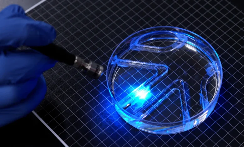
Researchers at the Massachusetts Institute of Technology (MIT) have successfully engineered a breakthrough in biohybrid robotics, demonstrating that an exceptionally thin layer of living muscle cells can generate sufficient force to propel a micro-robot through an aquatic environment. Bypassing the need for bulky motors, heavy gearboxes, or rigid mechanical actuators, this novel device represents a major leap forward in the integration of biological tissues with synthetic frameworks.
Published in the academic journal Advanced Functional Materials, the study details the creation of a swimming micro-robot that matches the approximate length and width of a standard stick of chewing gum. The device features a flexible, two-dimensional gel structure split into two distinct fins. Each fin is coated with a layer of genetically modified mammalian muscle cells significantly thinner than a human hair. By shining light onto these engineered tissues, researchers can trigger targeted muscle contractions, causing the fins to flap and drive the robot through water with precise navigational control.
The achievement marks the first time a two-dimensional, micro-scale biohybrid robot has achieved untethered locomotion using living muscle tissue in this manner. Experts believe the breakthrough could pave the way for a new generation of soft, responsive, and environmentally adaptable machines designed for delicate ecological monitoring and underwater exploration.
Main Facts of the Biohybrid Innovation
Traditional robotics relies heavily on synthetic materials, electric motors, and rigid actuators. While effective for industrial applications, these hard components can be hazardous, intrusive, or inefficient when operating in delicate natural habitats or interacting with fragile biological structures. To address these limitations, biohybrid robotics combines living biological tissue—which is naturally soft, self-healing, and responsive to chemical and physical stimuli—with synthetic polymer scaffolding.
Until now, however, most biohybrid robots have been constrained by three-dimensional designs. These legacy devices typically utilize bulky, lab-grown skeletal muscle chunks requiring millions of individual cells to manufacture. The MIT research team, led by Ritu Raman, associate professor of mechanical engineering, set out to determine whether a much simpler, ultra-thin 2D architecture could deliver comparable or superior mechanical performance.
The resulting robot consists of a gelatin methacrylate (GelMA) film measuring approximately half a millimeter in thickness. This precise dimension provides an optimal structural balance: it is sturdy enough to support a continuous layer of living cells while remaining lightweight enough to prevent the contracting muscle tissue from delaminating or tearing away from the base during operation.
The muscle cells coating the fins were genetically engineered to contract instantly upon exposure to visible light. By selectively illuminating the left fin, the right fin, or both simultaneously, the research team can dictate the robot’s heading, velocity, and turning radius. In experimental trials, the biohybrid swimmer successfully navigated an underwater maze inside a standard laboratory petri dish, covering a maximum distance of approximately four body lengths per minute.
Chronology and Development History
The creation of the swimming micro-robot did not happen in a vacuum; it is the culmination of years of methodical experimentation in biofabrication and tissue engineering at MIT.
The project builds directly upon foundational work conducted in Raman’s laboratory during the preceding year. At that time, the research group engineered an artificial muscle disk inspired by the mechanics of the human iris. For that experiment, the engineers patterned a soft fibrin gel disk with microscopic surface grooves radiating from the center like concentric rings. Living muscle cells were seeded onto this grooved surface, where they successfully organized into a thin, functional layer. When illuminated, the muscle tissue contracted multidirectionally, causing the disk to expand and compress in a manner mimicking the human pupil.
While the artificial iris successfully demonstrated that ultra-thin muscle layers could be induced to move in complex ways, the scale of movement was severely limited. The cells displaced themselves by a mere 100 microns—a microscopic distance that offered little practical value from a macroscopic robotics perspective. Furthermore, the fibrin gel used as the foundational substrate proved to be excessively soft, causing the material to warp and shrink under the mechanical tension exerted by the contracting muscle fibers.
Recognizing these limitations, the MIT team initiated a rigorous optimization phase to enhance both the force output and the physical displacement of the engineered muscle tissue. The researchers systematically evaluated three critical parameters of the hydrogel foundation: its chemical formulation, its structural stiffness, and the geometry of the surface micro-grooves designed to guide cellular alignment.
Through systematic trial and error, the engineers tested various groove architectures, comparing narrow channels with square profiles against longer channels featuring curved bottoms. They discovered that muscle cells aligned significantly better within square-bottomed grooves. This cellular alignment is crucial because muscle cells that grow in parallel are far more likely to fuse together into cohesive, multi-nucleated muscle fibers capable of generating synchronized, high-magnitude contractions.
Additionally, the team replaced the vulnerable fibrin gel with gelatin methacrylate (GelMA), a biocompatible polymer widely utilized in tissue engineering. By tuning the chemical cross-linking density of the GelMA, the researchers fabricated substrates with varying degrees of stiffness. They observed that firmer gels not only supported better cellular alignment but also enabled the muscle tissue to exert significantly greater mechanical force without degrading the underlying structure.
Prior to integrating the muscle-laden gel films into the swimming robot, the team subjected the biohybrid tissues to a regimen of physical conditioning resembling exercise. By exposing the engineered muscle to repeated, pulsed light stimulation, the cells were forced to contract rhythmically, strengthening the tissue and optimizing its responsiveness before deployment in aquatic trials.
Supporting Data and Performance Metrics
Evaluating the performance of micro-scale aquatic robots requires contextualizing their speed and mechanics against both biological organisms and traditional synthetic devices. Moving through water presents severe hydrodynamic challenges compared to locomotion in air, primarily due to the high density and viscosity of aqueous fluids relative to gaseous environments.
At its peak performance, the MIT swimming robot achieved a velocity of roughly four body lengths per minute. For comparison, an elite human Olympic swimmer can travel up to 65 body lengths per minute under optimal conditions. However, the biohybrid robot’s relative speed places it on par with certain slow-moving marine species, such as the cow shark, which cruises leisurely through ocean currents.
Given its microscopic dimensions and the absence of a heavy onboard power source or motor, the force output of the half-millimeter-thick muscle layer is exceptionally high. "It takes a lot of force to move through water versus air," notes Ritu Raman. "The robot’s quite strong, given its size."
The project received partial financial backing from the Office of Naval Research, reflecting the United States military’s ongoing interest in biomimetic systems capable of silent, efficient propulsion and stealthy aquatic navigation.
Official Responses and Expert Perspectives
The research team emphasizes that the successful navigation of the underwater maze serves primarily as a proof-of-concept rather than the final iteration of the technology. The primary scientific objective was to demonstrate that a two-dimensional layer of engineered biological tissue could be harnessed to generate directional, propulsive force in a fluid medium.
According to Maheera Bawa, the study’s first author, the integration of living tissue into mechanical designs opens entirely new avenues for how robots interact with their environment. The research paper, co-authored by MIT scientists Arielle Berman, Laura Schwendeman, Ferdows Afghah, and Seanbiron Johnson, highlights the distinct advantages of biohybrid architectures over conventional hard-bodied hardware.
"We believe that biohybrid robots powered by living muscle could one day perform delicate jobs like exploring environments too fragile or unpredictable for conventional hardware," Raman explains. Because living tissue is inherently compliant, soft, and sensitive to external stimuli, it minimizes the risk of damaging delicate ecosystems during data collection. Furthermore, biological components possess intrinsic self-healing properties that could theoretically allow autonomous repair mechanisms in remote or hazardous operating conditions.
Broader Impact and Future Implications
The development of ultra-thin, muscle-powered swimming robots signals a paradigm shift in soft robotics and bioengineering. By transitioning away from bulky, three-dimensional muscle masses toward highly efficient, two-dimensional functional layers, engineers can drastically reduce the material and economic costs associated with cultivating mammalian cells in laboratory environments.
The implications of this research extend far beyond academic demonstrations in controlled laboratory dishes. Future iterations of the technology could be deployed in aquatic ecosystems for continuous environmental monitoring, water quality testing, and ecological surveillance in fragile marine habitats such as coral reefs, where rigid metallic or plastic autonomous underwater vehicles (AUVs) risk causing physical abrasion or pollution.
Looking ahead, the MIT team has already established clear engineering objectives for the next phase of development. The primary focus will be optimizing the morphological design of the robot’s body and fins to hydrodynamic drag reduction, thereby enabling significantly higher swimming speeds.
"Our next goal is to optimize the body design to enable faster swimming," concludes Raman. "But even at slow swim speeds, one could imagine a muscle-powered swimmer being used for purposes like environmental monitoring in aquatic environments." As researchers continue to refine the synergy between living biology and synthetic engineering, the boundary between living organisms and machines continues to blur, promising a future where robots are grown rather than manufactured.